While scientifically, human cryopreservation is a compelling vision, scientists, entrepreneurs, and a growing cryonics community are believing in this vision, turning it into a legitimate reality. Many companies have already started offering “human cryopreservation” services. Here is how they do it:
As soon as the person is legally dead, the process of freezing must start almost immediately, else the cells enter a decomposition phase. First, blood is stabilized with a heart-lung resuscitator to supply the brain with oxygen; dry ice surrounds the body to help start cooling it gradually; finally, blood thinners are injected into the body to prevent blood coagulation.
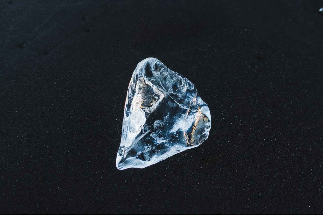
To understand how cryopreservation works, first we must understand what it means. The word “cryopreservation” can be divided into “cryo” and “preservation.” Cryo- is a prefix meaning “cold.” Cryobiology is the branch of science that studies the effects of very low temperatures on organisms.
At low temperatures, biological functions and chemical reactions are entirely halted. This is because, at very low temperatures, the proteins are not destroyed but rather frozen. This process is reversible, meaning that when the temperature rises to normal levels again, the protein will thaw and resume its functions, whereas at hot temperatures, proteins are irreversibly destroyed.
Cryopreservation is the process of preserving cells, tissues, organs, and other biological constructs using very low temperatures, i.e., -150 to -273 °C. In cryopreservation, reducing temperatures lowers the chemical reactions to an almost full stop. The preserved entities are maintained at such temperatures and then thawed when needed for use. For example, egg and sperm banks cryopreserve eggs and sperm until you are ready to use them.
So, can humans be cryopreserved and revived after 10, 100, even 1000 years? The truth is that this is highly challenging and scientists are pursuing different effective approaches to reviving tissues after cryopreservation.

Cryopreservation is a delicate process that does not guarantee the survival of all cells. Cells are very delicate and highly affected by their environment. In fact, several factors can prevent the revival of cells after cryopreservation, such as ice and crystal formation, improper thawing, and osmotic shocks. Additionally, while single cell suspensions (eggs or sperms) are easy to handle, tissues containing billions of cells are another level of complexity.
Another challenge is the speed of thawing. Fast cooling can cause cell injuries while slow cooling can cause what is known as osmotic shocks (increased internal pressure as water rapidly enters the cells) which can lead to cell death. Rapid cooling and thawing in equal distribution is a challenge scientists are facing to find the optimal methods of preservation.
Scientific origin of cryopreservation
The recorded history of cryopreservation dates back to the late 1800s when it was used to preserve both sperm and red blood cells. In 1953, an early theoretician of cryopreservation, James Lovelock, discovered that the cryopreservation process caused osmotic stress in cells. He flash-froze the liquid containing the cells and noticed the formation of ice crystals in the red blood cells. In 1954, the first human insemination experiment with previously frozen sperms was successful and resulted in three pregnancies.
Science has come a long way since then. Cryopreservation has substantially improved, offering hope for a better tomorrow.
Human cryopreservation process
The blood is replaced with cryoprotectants (CPA) known as the body’s antifreeze. CPAs are injected to prevent ice formation. Once the cryoprotectants reach the equilibrium amount, the cells are cooled down at a rate of 1 °C/min, until a temperature of -130 °C is reached.
The body is then stored head down, to protect the head in the event of a leak, in a metallic tank called a dewar, filled with liquid nitrogen at a temperature around -196 °C. The patient must then await a medical breakthrough to eventually rise and shine.
The good news is that no electricity is required in cryopreservation since the body is stored in liquid nitrogen, and you do not have to worry about the electric bill. But, the costs of cryonics range anywhere between $28,000 up to $200,000.
Applications of cryopreservation
There are many successful accomplishments using cryopreservation.
Embryo preservation with the application of IVF cycles has been successful since 1984. Fertility preservation for eggs and sperm, especially for patients undergoing chemotherapy and radiation therapy, has given hope to patients with cancer and other illnesses.
According to The Hewitt Fertility Centre in Liverpool, the survival rates are approximately 90-95%. So survival itself would not be enough to allow for a safe transfer of the thawed embryo. The embryo would need to show signs of life when it starts to re-expand in a process that could take up to 2 hours.
Stem cells are also currently cryopreserved. They are a new elixir in medicinal therapies, especially in the treatment of spinal cord injuries, type 1 diabetes, Parkinson’s, Alzheimer’s, cardiovascular diseases, strokes, burns, and cancer.

Another example of successful cryopreservation is that of hepatocytes, the cells of the liver. They help detoxify the body, break proteins, and boost metabolism and immunity.
Additionally, blood and organs are also cryopreserved.
You may be surprised to know that there is currently a “seed bank” in which the seeds of thousands of plant species are cryopreserved, in case the earth is destroyed.
An honorable mention is for the Siberian bdelloid, the tiny worm-like animal that was recently discovered to be naturally cryopreserved for 24,000 years, which broke all the odds.
The latest technique of thawing cryopreserved organs
Before delving into the topic of cryopreservation and the thawing process, we must start with reviewing the latest published nanotechnology protocols, known as nanowarming, to sufficiently thaw cryopreserved large tissues (≥3 mL) and organs.
Nanowarming has been used for the first time to rewarm a rat kidney rapidly and uniformly. In addition, studies have shown that the process has been carried out successfully, free of both ice crystallization and cracking. The protocol applied in the process consists of the following three steps:
- Perfusion: The organs are supplied with the cryoprotective cocktail (CPA) and silica-coated iron oxide nanoparticles (sIONPs) through their renal artery.
- Verification: To ensure that the sIONPs are uniformly distributed in the vitrified system, the organ is cooled at -40 °C/min in a controlled rate freezer. Then a microcomputed tomography (µCT) imaging is used to verify the distribution of sIONPs.
- Nanowarming: The distributed sIONPs are then excited by applying a radiofrequency field (RF), causing a nanowarming process at a mean rate of 63.7 °C/min. Meanwhile, another study assumes that the nanowarming technique could potentially uniformly thaw organs at 100 °C/min.
The protocol of thawing cryopreserved stem cells
Another thawing protocol is used for the thawing of human embryonic stem (hES) cells. We have summarized these nine steps that are only meant to be carried out by professionals in regulated facilities, so don’t try this at home!
- Pre-experimental setup: In this step, and one day before the thawing, a thin feeder layer is placed on the cells. The feeder consists of irradiated mouse embryonic fibroblasts (MEF), CF1 mouse strain, on one well of a gelatin-coated 6-well tissue culture plate in MEF culture medium. That is then incubated overnight at 37°C and 5% CO2.
- Defrosting: This starts with removing a cryogenic vial of the cells from the liquid nitrogen storage tank using metal forceps. Then, the frost is removed by rolling the vial between gloved hands for 3-5 seconds.
- Bathing: In this step, the vial is immersed and swirled gently into a 37°C water bath. During the process, the vial needs to be closely checked to see the size of the ice crystals. During bathing, you should not submerge the cap of the vial in order to avoid contaminating the cells.
- Sterilization: The process shall be performed in a sterile biological safety cabinet. Only if a small ice crystal remains, submerge the entire vial in ethanol to sterilize. Otherwise, transfer the vial contents directly to the bottom of a 15 mL conical tube.
- Centrifugation: Begin with slowly adding 4 mL of cell culture medium to the tube and swing gently to mix the cells as the new medium is added to the tube. The process will last 5 minutes at 200 x g.
- Plating: After the centrifugation is completed, the pelleted cells are brought back to the sterilized cabinet. The supernatant is carefully aspirated without aspirating the cell pellet. The pellet is then resuspended gently by adding 2.5 mL of hES cell culture medium using a 5 mL glass pipet and pipetting 3-4 times.
- Well preparation: The MEF feeder from step 1 is aspirated, and 1.0 ml of PBS (phosphate-buffered saline) is added to the well. The PBS is then aspirated while adding all 2.5 mL of the hES cell suspension to the prepared well of the 6-well plate.
- Incubator: The plate is placed into the 37°C incubator and carefully slid backwards and forwards, from side to side, to evenly distribute the cells throughout the well. The cells will need to attach at 37°C and 5% CO2 overnight.
- After the thaw: The medium is removed on the following day, and 2.5 ml of fresh hES cell culture medium is added to the well.
Conclusion
Cryopreservation has undoubtedly been a blessing to humans so far. Through cryopreservation, science has offered us a technique to preserve human cells and organs at low temperatures, giving it a chance of a safe future thawing.
Scientists have sought cryopreservation since the nineteenth century, and the process has evolved over the years. The cryonics community is still pushing, and there is no limit to what science can make possible. The cryopreservation procedure is becoming a commercial standard, a business model, and a scientific goal for researchers and investors worldwide.
We can already observe the progress accomplished in thawing techniques. In addition, several studies have shown promising results in the uniform and sufficient revival of vitrified stem cells and organs. Applying nanowarming to the thawing of cryopreserved large tissues and organs has proven effective at a rapid rate of up to 100 °C/min. Such progress in cryopreservation brings great hopes not only for the field of organ transplantation but also for full-body options or at least neuropreservation options awaited by the cryonics community.
We are still waiting for more breakthroughs to uncover how cryopreservation works and how it can even be optimized to offer better chances of revival for cryopreserved humans.