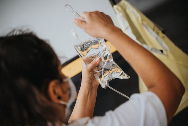
Cryonics involves freezing down tissues, organs – or more sanguinely the whole body – of an organism so that they become metabolically inert and in a dormant stage that would theoretically make it possible for scientists or researchers to revive the tissue and restore its function later on in the future. However, extremely low temperatures (-120 degrees or even less) are usually needed to attain such molecular inactivity inside the cells, causing substantial damage to the cells and tissues that make it harder to re-establish the functional properties upon thawing.
What causes damage?
The associated damage is generally caused by two different mechanisms. Firstly, during the freezing process, where ice crystals form inside the cells due to the high water content. This causes mechanical damage as the shape of the cells is distorted by these crystals often causing rupturing and thus displacing the cells. Damage is also caused by the chemical and osmotic effects of solutes concentrated in the unfrozen residual water between the formed crystals, often known as “solution effects” injury. The damage can be minimized by careful modulation of temperatures, and by using different cryoprotectants.
Using cryoprotectants for damage control during freezing
Any chemical that is sufficiently soluble in water and has a melting point lower than zero degrees celsius can be used as a protective agent during the process of freezing tissues or organs. In order for a solution to be a good cryoprotectant, it needs to effectively depress the melting point of water, must not precipitate or form hydrates, and should be non-toxic to cells even at high concentration. 5-15% of cryoprotectant is usually enough to allow isolated cells to survive after freezing or thawing at liquid nitrogen temperatures. Cryoprotectants reduce the damage to the cells as their presence creates large pockets of unfrozen liquid. This reduces the impact that would otherwise be caused by the growing ice crystals.

Usually, there are two classes of cryoprotectants: penetrating and non-penetrating. The size of the molecules of non-penetrating cryoprotectants, for example polyethylene glycol (PEG), are larger and hence cannot penetrate the cells. Penetrating cryoprotectants, such as glycerol and dimethyl sulfoxide (DMSO), are the preferred type and have a relatively smaller molecular size. They are able to enter the cells and remain there, hence preventing excessive dehydration of cells during the freezing process. Cryoprotectants are important because they influence the physical properties of the intracellular solution, reduce intracellular ice formation, increase resistance to cold shock, help maintain the integrity of protein and lipid structure of the cell membrane which increases the membrane’s fluidity. While the conventional freezing technique works to an acceptable level in cell suspension, if our aim is to preserve larger tissue or organ samples, there is always a risk of physical disruption, like hairline cracks. But not only during the freezing process, a series of complexities can arise due to temperature fluctuations during the thawing process.
Vitrification to avoid ice formation altogether
To avoid such damage during revival, vitrification as an alternative to freezing has been empirically proven to be a superior technique for cryopreservation of tissues and organs. Proposed for the first time in 1984 by cryobiologist Gregory Fahy, the term vitrification means to “turn into glass”. This approach involves cooling the samples so fast that the formation of ice crystals is avoided, and turning the sample into a steady, non-crystalline amorphous state. To achieve successful vitrification, the tissue or organ should be loaded with just enough cryoprotectant to prevent ice from forming during the rapid cooling process; with a significantly faster rate of cooling, the samples can be vitrified with a lesser concentration of cryoprotectant as compared to the conventional freezing approach.
Specially formulated cryopreservation solutions are used in vitrification procedures. Usually, the solutions contain a cryoprotectant along with another carrier solution. The carrier solution is not the primary cryoprotectant per se, but a mixture of salts, osmotic agents, pH buffers and nutritive ingredients, as well as apoptosis inhibitors. One of the most widely used vitrification solutions is called VS55, which contains DMSO, formamide, and propylene glycol as its ingredients.

For vitrification and subsequent revival of vitrified samples, rates of cooling and warming at 10° and 80°C/min respectively, have been deemed to work considerably well. Though this rate of cooling is feasible in smaller volumes (≤3 ml), preserving larger tissues and organs is more complex. Using convective warming alone to revive any vitrified sample is highly inefficient due to devitrification and/or cracking. The advances made in vitrification (rapid cooling) have not been matched by technologies that are sufficiently efficient in rewarming/thawing. The critical warming rate (CWR) required to avoid devitrification – the process of crystallisation during warming – is higher than the corresponding critical cooling rates (CCR). For a reproducible and successful thawing, these rates need to be uniform throughout the material so as to avoid thermal gradients that cause thermal stress, producing fractures and cracks. For larger volumes, it is almost impossible to generate such uniform rates. Hence, convective warming is rendered useless.
Nanowarming as a novel approach for damage-free revival
The developments in nanotechnology have shown that it indeed is possible to revive the functional properties of cryopreserved tissues of even larger volumes. The rewarming problem was solved by a group of scientists at the University of Minnesota by using nanoparticles. They used silica-coated iron oxide nanoparticles (msIONP) in a cryoprotectant solution (VS55). Inductive activation of nanoparticles was done by using noninvasive electromagnetic (Radio Frequency) waves. The electromagnetically activated nanoparticles act as tiny heaters around the tissues, hence achieving a uniform and sustained rate of heating in the range of 100-200 degrees celsius per minute. When the tissues were examined for viability post-revival, they did not show any damage as compared to control samples that were conventionally rewarmed. The group showed that tissues as diverse as porcine carotid arteries, porcine heart valves, and femoral arteries, up to 80 ml in volume, can be metabolically revived using this approach.
Hence, in the future when this new nanotech is more advanced, we can expect to revive even larger volumes of tissue without damage, and hopefully even entire bodies.